

Wound

Introduction
Wound is discontinuity or break in surface epithelium
Simple when only skin is involved
Complex when it involves underlying nerves, vessels and tendons
Types of wounds
Closed Wound
Contusion
Minor soft tissue injury without break in skin
It produces discoloration of skin due to collection of blood underneath

Abrasion
Epidermis of skin is scraped away exposing dermis
Painful, as dermal nerve endings are exposed

Hematoma
Collection of blood following injury
It can be subcutaneous, intramuscular, subperiosteal
They get absorbed, if not they get infected

Open Wound
Incised Wound
Caused by sharp objects
Has sharp edge and it is less contaminated
Primary suturing is ideal

Lacerated Wound
Caused by blunt injuries
Due to blunt nature of object, there is crushing of tissue which may result in hematoma, bruising or even necrosis of tissue

Penetrating wound
Look like innocent injury with small, 1 or 2 cm long cut but internal organs such as intestines, liver, spleen or mesenteric blood vessels may have been damaged

Crushed or Contused wounds
Caused by blunt trauma
May cause severe hemorrhage, death of tissue and crushing of blood vessels.
More prone to gas gangrene, tetanus

Tidy Wounds
Incised, clean, healthy tissue
Untidy Wounds
Crushed or avulsed, contaminated, devitalised tissue and often with tissue loss
Acute Wound
Stab wound, blast injuries, RTA
Chronic Wound
Leg ulcers, pressure sore
Healing of wound
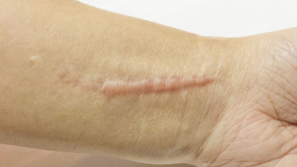
By primary intention- Produces clean, neat, thin scar
By secondary intention- Produces ugly scar
Components of wound healing
Inflammatory phase (Lag Phase)
Release of mediators of inflammation like histamine from platelets, mast cells and granulocytes. This causes increase capillary permeability
Later kinins and prostaglandins act and they play a chemotactic role for white cells and fibroblasts
In first 48 hours, polymorphonuclear leukocytes dominate. They play role of scavengers by removing dead and necrotic tissue
Proliferative Phase (Collagen Phase)
Between 3rd and 5th day, polymorphonuclear leukocytes diminish and monocytes increase. They are specialized scavengers
By 5th and 6th day, fibroblast appear, proliferate and eventually give rise to protocollagen which is converted in collagen
Enzyme protocollagen hydroxylase, oxygen, ferrous ions and ascorbic acid are necessary for this step
Epithelialisation occurs from edges of wound by process of cell migration and cell multiplication by marginal basal cells. Within 4 hours entire wound is re-epithelialised
Remodelling Phase (Maturation)
Starts after 4th day and completed by 14th day
Brought by specialised fibroblast called as myofibroblasts
Corticosteroids, irradiation, chemotherapy delay wound contraction
Phase of Scar formation
Fibroplasia and laying of collagen is increased
Vascularity become less
Epithelialisation continues
Ingrowth of lymphatics and nerve fibres takes place
Remodelling of collagen takes place with cicatrisation, resulting in a scar
Complication of wound healing
Infection- Majority of bacteria are endogenous
Ugly scar
Keloid and hypertrophic scar
Incisional Hernia and wound dehiscence
Pigmentation
Marjolin's ulcer
Wound closure or wound suturing
Primary suturing-
Within few hours
It should be incised or cut wound
There should not be any infection
Foreign body if present should be removed
Associated injuries to other structure should be repaired
Injection TT should be given
Wound excision and primary suturing
When wounds edges are jagged
Wound is contaminated with organisms or foreign body
Tissue are crushed
In such conditions, wound is explored and devitalised tissues and foreign body if present are removed. The wound is irrigated with antiseptic agents.
Precautions
Done within 6 hours
Tetanus and gas gangrene prophylaxis
Repair of tendons and nerves can be done at later date if contamination is excessive
Wound excision and delayed primary suturing
Indicated in lacerated wounds with major crush injuries.
Primary suturing within 6 hours is not done in these wounds because of
Gross oedema
Increased tissue tension
Hematoma
Contamination with bacteria
Principles of debridement
Done under General Anaesthesia
Assess the extent of injuries and loss of tissue
Control bleeding
Excision of devitalised tissue
Good saline wash than betadine and hydrogen peroxide
Complication of skin loss
Secondary infection
Underlying structures are in danger
Septicemia
Deformity and disability in later date
Secondary Suturing
Suturing after 7 to 14 days later after controlling infection
Skin is freed from the edges of the wound and the granulation tissue and skin are approximated
Factors affecting wound healing
General factors
Age
Dermal collagen content decrease with aging
Collagen fibres show distorted architecture and organisation on aging
Malnutrition
Delayed because of deficiency of vit C
It is important for synthesis and maintenance of collagen
Following injury vitamin C deficiency can occur after 3 to 4 weeks
Zinc deficiency is known to delay the healing of pilonidal sinus
Albumin levels must be less than 2 g/dL to have an effect on wound healing
Diabetic Patient
Due to microangiopathy, atherosclerosis, decreased phagocytic activity and proliferation of bacteria
Jaundice and uraemic patient
Fibroblastic repair is delayed
Cytotoxic drug like doxorubicin
Malignancy
Chemotherapeutic drug causes
Decreased mesenchymal cell proliferation
Reduce number of platelets
Reduced inflammatory cells
Reduce growth factors
Decrease wound breaking strength
Generalised infection
Corticosteroids
If given early after healing they do no interfere
Local Factor
Poor blood supply
Wound on knee and shin of tibia
Local infection
If bacteria count exceeds 10,0000 organism/ mg and if beta hemolytic streptococci are present then wound does not heal
Collagen synthesis is reduced and collagenolysis is increased
Hematoma
Faulty technique
Tension
Hypoxia
Killing property of macrophages reduced
Production of fibroblasts reduced
Collagen synthesis affected
Smoking causes vasoconstriction and elevate carbon monoxide level
Ionizing radiation
Hypertrophic Scar
Hypertrophy of mature fibroblasts in hypertrophic scar.
Occurs from prolonged inflammatory phase
No itching
Non tender
Not vascular
Does no extend beyond the boundary of original incision or wound
Precipitating Factor
Over Sternum
Over Joints
Young person
Treatment
Often not necessary
Stocking, gloves
Elastic bandage
Excision can be done
Topical retinoids
Keloids

In keloid, proliferation of immature fibroblasts with immature blood vessels are found
Collagen bundle are virtually absent
Marked tendency for local recurrence after excision
Common site- Over Sternum
Continue to get worse even after 1 year
Severe itching
Margin is tender
Vascular red and erythematous
Precipitating factors
Generally in black races
Tuberculosis patient
Hereditary
Vaccination sites
Does not become small
Complication
Ulceration
Infection
Treatments
Injection Kanacort
Intrakeloidal excision and skin grafting
Silicone application
Topical retinoids
Keloid Sites
High chances
Skin of ear lobe
Presternal
Deltoid
Upper back
Least Chances
Eyelid
Genitalia
Palm
Sole
Across joint
Response to injury
Depends upon severity of injury
Neuroendocrine response
Earliest to get activated
Starts with nociceptive receptors
Stimulation of hypothalamus leads to release of corticotrophin releasing hormone (CRH), which triggers release of ACTH from anterior pituitary.
ACTH act on adrenals to increase the secretion of cortisol.
Activation of sympathetic nervous system, which in turn causes release of adrenaline and glucagon
Increased release of growth hormone and glucagon

Hormones which increase following injury
ACTH
GH
Glucagon
Cortisol
Adrenaline
Hormone whose level fall following inury
Insulin
Thyroid hormones
Testosterone
Immune response
Pro inflammatory response
Causes
Pyrexia, due to action of pro inflammatory cytokines on hypothalamus
Proteolysis in skeletal muscles
Production of acute phase protein in liver
Pro inflammatory cytokines
IL- 1
IL- 6
IL- 8
TNF alpha
Anti inflammatory response
Anti inflammatory cytokines
IL- 4
IL- 5
IL- 9
IL- 13
TNF Beta
Comments